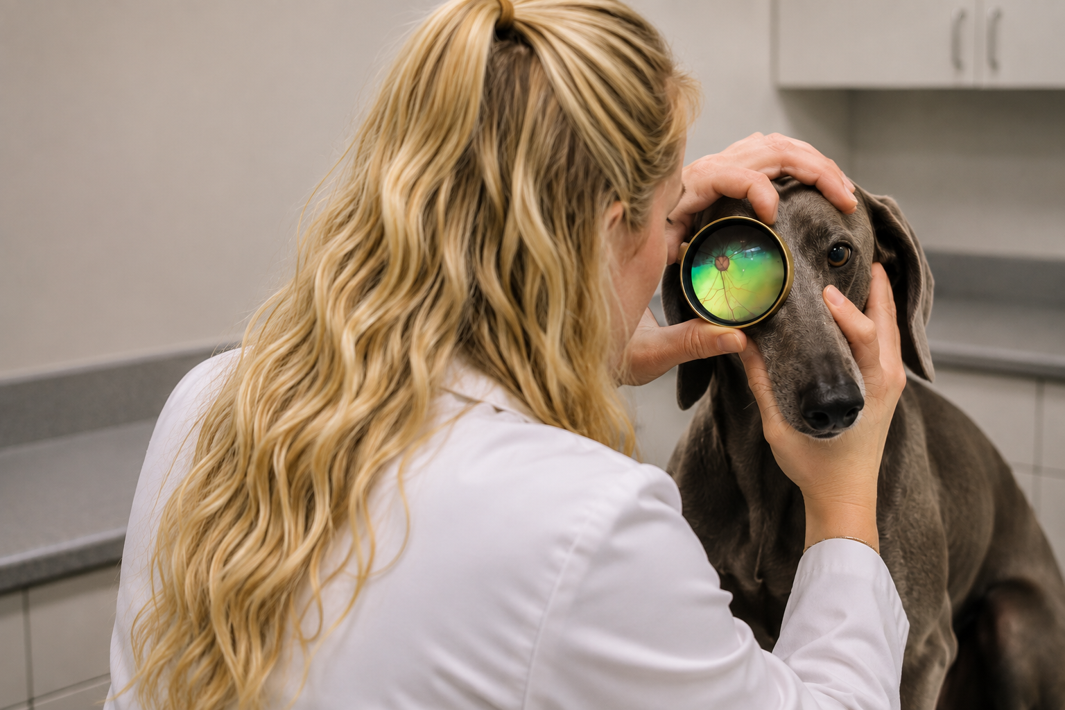
Retinal imaging gives our veterinary team a clearer view of structures that may be difficult to fully assess during a routine eye examination. These detailed images can help us identify abnormalities, document changes, and determine when additional evaluation or treatment may be appropriate.
If you have noticed changes in your pet’s vision, eye appearance, or behavior—or simply want to learn more about advanced veterinary eye care—our team is here to help.
What Is Veterinary Retinal Imaging?
Retinal imaging is a diagnostic technique used to capture detailed images of the retina and other structures located at the back of the eye. The retina is a thin layer of light-sensitive tissue that plays an essential role in vision.
During a traditional veterinary eye examination, your veterinarian may examine these structures using specialized instruments. Digital retinal imaging enhances this evaluation by creating images that can be reviewed, documented, and compared over time.
At FurBaby Medical Veterinary Practice LLC, the Volk VistaView™ helps our veterinary team obtain high-quality retinal images as part of a thorough ocular assessment when clinically appropriate.
What Can the Volk VistaView™ Help Us See?
The Volk VistaView™ allows veterinarians to capture images of important structures within the back of the eye, including the retina and optic nerve region.
These images can provide valuable information about changes that may be associated with retinal disease, inflammation, trauma, systemic health conditions, or other ocular abnormalities.
Retinal imaging does not replace a complete veterinary examination. Instead, it provides additional diagnostic information that can help your veterinarian better understand what may be happening inside your pet’s eyes.
Why Is Retinal Health Important for Dogs and Cats?
Healthy retinas are essential for normal vision. When retinal disease develops, pets may experience subtle vision changes that are easy to overlook at first.
Dogs and cats cannot tell us when their vision becomes blurry or when they begin having difficulty seeing in certain lighting conditions. Instead, pet owners may notice behavioral changes.
Signs Your Pet May Be Having Vision Problems
Contact your veterinarian if your pet begins:
- Bumping into furniture, walls, or other objects
- Hesitating before using stairs
- Having difficulty navigating unfamiliar environments
- Becoming reluctant to go outside at night
- Appearing disoriented in dim lighting
- Becoming unusually startled when approached
- Having trouble locating toys, treats, or food
- Developing unusual redness, cloudiness, or changes in eye appearance
- Showing differences in pupil size or response to light
Sudden vision loss or significant eye pain should always be evaluated promptly.
What Conditions Can Retinal Imaging Help Evaluate?
Retinal changes can occur for many different reasons. Depending on your pet’s symptoms, medical history, and examination findings, retinal imaging may assist your veterinarian in evaluating or documenting abnormalities involving the back of the eye.
Retinal Degeneration
Some dogs and cats can develop degenerative retinal diseases that gradually affect their ability to see. Imaging may help document the appearance of the retina and support further diagnostic evaluation.
Retinal Detachment
A retinal detachment occurs when the retina separates from its normal position. This can cause significant or sudden vision problems and may be associated with underlying ocular or systemic disease.
Inflammation
Inflammatory conditions can affect structures inside the eye. Detailed visualization can help your veterinarian recognize abnormalities and determine whether further testing or referral is needed.
Changes Associated With Systemic Disease
The eyes can sometimes provide clues about conditions affecting other parts of the body. Certain systemic illnesses, including high blood pressure and some infectious, inflammatory, or metabolic diseases, may cause changes that can be observed during an ocular examination.
Trauma
Eye injuries can sometimes affect structures that are not easily visible from the outside. A thorough veterinary eye examination can help determine the extent of an injury and whether deeper ocular structures may be involved.
Optic Nerve and Retinal Abnormalities
Retinal imaging may also help document unusual changes involving the optic nerve region or surrounding retinal tissue.
Benefits of Digital Retinal Imaging for Pets
One of the biggest advantages of digital retinal imaging is the ability to create a visual record of your pet’s eye health.
Detailed Visualization
Advanced imaging provides a detailed look at structures within the back of the eye, giving your veterinarian additional information beyond what can be seen externally.
Baseline Documentation
An image taken today can provide a valuable baseline for future comparison. If your pet develops changes later, your veterinarian may be able to compare new images with previous findings.
Monitoring Changes Over Time
For pets with known ocular abnormalities, repeat imaging may help document whether a condition appears stable or has changed.
Supporting Veterinary Decision-Making
Images can complement your pet’s physical and ocular examination findings and help your veterinarian determine whether additional diagnostic testing, treatment, monitoring, or specialist evaluation should be considered.
What Should I Expect During My Pet’s Retinal Imaging?
Your veterinarian will first evaluate your pet and determine whether retinal imaging is appropriate.
The exact process can vary depending on your pet’s temperament, eye condition, and reason for the examination. Your veterinary team will position your pet so images of the back of the eye can be obtained with the Volk VistaView™.
Your veterinarian can then review the images alongside the findings from your pet’s examination and medical history.
Does My Pet Need Retinal Imaging If Their Eyes Look Normal?
Not all eye conditions produce obvious external changes.
An eye may appear relatively normal to a pet owner even when changes are occurring internally. This is one reason veterinary examinations are important, particularly if your pet is experiencing changes in behavior or vision.
Your veterinarian can determine whether retinal imaging or additional eye testing would provide useful information based on your pet’s individual circumstances.
Retinal Imaging for Senior Pets
Vision changes become particularly important to monitor as pets age. Senior dogs and cats may develop ocular conditions alongside other age-related health concerns.
If an older pet starts hesitating on stairs, becoming uncomfortable in darkness, bumping into objects, or seeming less confident in unfamiliar surroundings, these changes should not automatically be dismissed as normal aging.
A veterinary examination can help determine whether vision loss, pain, cognitive changes, mobility problems, or another health condition may be contributing to the behavior.
Frequently Asked Questions About Retinal Imaging for Pets
Why would my veterinarian recommend retinal imaging?
Your veterinarian may recommend retinal imaging when additional visualization or documentation of the retina and other structures at the back of the eye could help evaluate your pet’s condition.
Can retinal imaging diagnose every eye condition?
No. Retinal imaging is one diagnostic tool and does not replace a complete veterinary eye examination or other diagnostic tests. Depending on the findings, additional testing or consultation with a veterinary ophthalmologist may be recommended.
Can retinal images be compared over time?
Yes. One benefit of digital imaging is the ability to document findings so they can potentially be compared with future images. This may be particularly helpful when monitoring known abnormalities.
Should I schedule an examination if my pet suddenly cannot see?
Yes. Sudden vision changes should be evaluated promptly. Contact a veterinarian as soon as possible if your pet suddenly begins bumping into objects, appears unable to see, develops significant eye redness or pain, or experiences another abrupt eye-related change.
Can eye changes be related to other health problems?
Yes. Changes within the eyes can sometimes be associated with systemic health conditions. Your veterinarian may recommend additional diagnostic testing if an eye examination raises concerns about your pet’s overall health.
Is retinal imaging only for older pets?
No. Although senior pets can experience age-related eye changes, retinal and ocular conditions can occur at many ages. Whether imaging is recommended depends on your pet’s symptoms, examination findings, medical history, and individual risk factors.
Advanced Veterinary Eye Care in Ware, MA
At FurBaby Medical Veterinary Practice LLC, we believe that advanced technology can provide valuable information when combined with attentive, individualized veterinary care.
The Volk VistaView™ retinal imaging system gives our veterinary team another way to examine and document important structures within your pet’s eyes. Whether your pet is experiencing possible vision changes or your veterinarian identifies an abnormality during an examination, retinal imaging can provide additional insight to guide the next steps in their care.
Contact FurBaby Medical Veterinary Practice LLC
If you are concerned about your dog or cat’s vision or would like to learn more about retinal imaging with the Volk VistaView™ in Ware, MA, contact FurBaby Medical Veterinary Practice LLC to schedule an appointment.
FurBaby Medical Veterinary Practice LLC
124 West St Ste F
Ware, MA 01082
Phone: (864) Furbaby (387-2229)